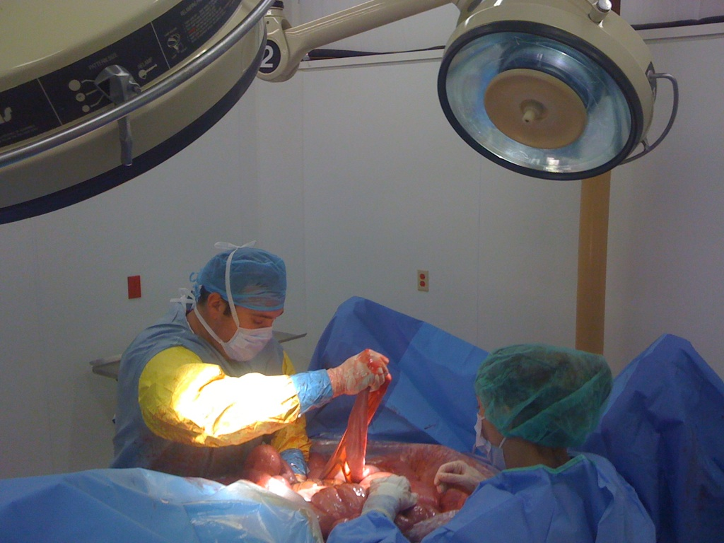
CALEC surgery, or Cultivated Autologous Limbal Epithelial Cells, represents a groundbreaking step forward in the treatment of corneal damage that was previously deemed untreatable. This innovative procedure utilizes cutting-edge stem cell therapy to not only restore the cornea’s surface but also significantly improves the quality of life for individuals suffering from severe eye injuries. During clinical trials, patients benefited from a remarkable success rate of over 90%, demonstrating CALEC’s efficacy as a viable alternative to traditional corneal transplants. By harvesting ocular stem cells from a healthy eye, surgeons are able to create a cellular graft that can regenerate the damaged areas of the cornea, offering new hope for those with vision impairment. As research continues, CALEC surgery may redefine eye injury treatment, opening doors for a range of corneal transplant alternatives that utilize a patient’s own biological materials.
The CALEC procedure, also known as cultivated limbal epithelial cell transplantation, is an innovative surgical approach aimed at restoring damaged corneal surfaces. Utilizing specialized stem cell techniques, this method offers an alternative to traditional corneal transplants, harnessing the body’s natural healing abilities. By extracting and expanding ocular stem cells from a viable eye, surgeons can create a graft that promotes the regeneration of the cornea, which is paramount for individuals afflicted by serious eye injuries. The success of CALEC surgery signals a promising advancement in ocular restoration therapies and provides a beacon of hope for those with chronic vision problems resulting from corneal damage. As the field of eye injury treatment evolves, alternative therapies like CALEC are expected to play a critical role in enhancing patient outcomes.
Understanding CALEC Surgery: A Revolutionary Approach to Corneal Restoration
CALEC surgery, or Cultivated Autologous Limbal Epithelial Cells surgery, represents a significant breakthrough in the field of ocular medicine. This innovative procedure utilizes stem cells harvested from a healthy eye to regenerate and restore the surface of a damaged cornea. During the process, limbal epithelial cells are extracted through a biopsy, expanded in a controlled laboratory environment, and then carefully transplanted into the affected eye. This state-of-the-art technique provides patients who were previously considered untreatable with a renewed hope for vision restoration and improved quality of life.
The results from clinical trials of CALEC surgery have been encouraging, demonstrating over 90 percent effectiveness in restoring the cornea’s surface. Such statistics indicate a remarkable advancement for individuals suffering from severe corneal injuries or limbal stem cell deficiencies caused by conditions like chemical burns and infections. Moreover, the careful coordination among multidisciplinary teams ensures the procedural safety and efficacy, making CALEC a promising alternative to traditional corneal transplants.
The Role of Stem Cell Therapy in Eye Injury Treatment
Stem cell therapy has emerged as a pioneering solution in the treatment of eye injuries, providing new avenues for healing in cases once deemed hopeless. Specifically, ocular stem cells play a critical role in the recovery of cornea surfaces by enabling the regeneration of vital tissues. The use of harvested limbal epithelial cells in the CALEC procedure is a perfect illustration of how stem cell therapy can provide functional and aesthetic improvements for patients suffering from debilitating eye conditions.
Furthermore, stem cell therapy goes beyond merely restoring vision; it also alleviates pain and discomfort associated with corneal damage. Many patients who participated in the clinical trial reported improved visual acuity and a significant reduction in symptoms. As the medical community continues to explore the applications of stem cell therapy, the future looks promising for innovative treatments that can transform how we approach eye injury treatment and corneal restoration.
Advancements in Corneal Transplant Alternatives
The traditional approach to treating severe corneal damage often involves corneal transplantation, a procedure that may not be viable for all patients, particularly those with limbal stem cell deficiencies. Corneal transplant alternatives, such as CALEC surgery, present a groundbreaking shift in treatment strategies. Instead of relying solely on donor tissue, CALEC utilizes the patient’s own stem cells, effectively eliminating the risk of transplant rejection while maximizing the chances of successful healing.
Additionally, the development of CALEC surgery opens doors to a wider range of patients who may not have had access to effective treatment options previously. With the potential for allogeneic grafts sourced from healthy donor eyes in future studies, CALEC might soon offer hope to individuals with bilateral corneal damage. By leveraging innovations, the field of ophthalmology is striving to ensure more patients can benefit from cutting-edge techniques that prioritize safety and effectiveness.
Clinical Trial Insights: CALEC’s Safe Application
The clinical trials conducted for CALEC surgery have highlighted not only its efficacy but also its impressive safety profile. In a comprehensive study, 14 patients with corneal damage underwent the procedure, with follow-up assessments showing substantial improvements in the corneal surface and visual acuity over 18 months. With no serious adverse events reported, the trials underscore the promise that CALEC holds as a safe treatment option for restoring vision and alleviating chronic pain stemming from corneal injuries.
Moreover, detailed follow-up data revealed that 93 percent of participants exhibited significant successes in corneal restoration at the 12-month mark. The overall performance indicates that CALEC surgery is not just a temporary fix but offers long-term benefits for eye health. This positive outcome paves the way for further research and development in stem cell-based therapies, potentially leading to long-lasting solutions for ocular disorders.
Potential Challenges of CALEC Surgery
Despite the promising results of CALEC surgery, certain challenges must be addressed before the procedure can be widely implemented. One of the primary limitations is the requirement for patients to have at least one healthy eye for the necessary biopsy collection. This requirement presents a significant barrier for individuals with bilateral corneal damage, necessitating the development of alternative grafting methods that utilize stem cells from donor sources.
Addressing these limitations will be vital for translating the success of CALEC surgery into broader applications. Ongoing research aims to explore allogeneic manufacturing processes, which will potentially enable the treatment of patients regardless of their individual circumstances. By overcoming these challenges, the ophthalmic community aspires to expand access to innovative stem cell therapies, creating equitable opportunities for all who suffer from debilitating eye injuries.
The Future of Ocular Stem Cells in Eye Treatments
The future of ocular stem cells in eye treatments is bright and full of potential. As the research community delves further into stem cell therapies, the advancements in understanding limbal epithelial cells and their regenerative capabilities are expected to unlock new treatment possibilities. The emphasis on innovative approaches, such as CALEC surgery, demonstrates a shift from traditional reconstruction methods to biologically-based solutions that harness the body’s own healing processes.
Ongoing trials and research will likely refine the techniques, allowing more patients to benefit from cutting-edge therapies. As our knowledge about ocular stem cells expands, we can anticipate a transformative impact on how we manage eye injuries and diseases, leading to improved outcomes for countless individuals suffering from visual impairments.
Collaborative Efforts in Stem Cell Research
Collaboration is at the heart of advancing stem cell research and its application in eye care. The successful development and implementation of CALEC surgery involved a concerted effort among various institutions, including Mass Eye and Ear, Dana-Farber, and Boston Children’s Hospital. Such partnerships foster innovation and ensure rigorous scientific evaluation, enabling researchers to overcome complex challenges in the pursuit of effective therapies.
Additionally, as more hospitals and research centers recognize the potential of stem cell therapies, the incorporation of multidisciplinary teams will be essential to elevate the standard of care for ocular patients. Their collective expertise can drive research forward, leading to significant breakthroughs that will refine current treatments and explore new possibilities for restoring vision and alleviating suffering.
Exploring the Clinical Application of CALEC Therapy
The clinical application of CALEC therapy signals a significant step forward in managing severe ocular conditions. By enabling the regeneration of the corneal surface through the unique method of cultivating stem cells, CALEC opens new avenues for treatment that are both innovative and patient-centered. The initial success of the therapy in clinical trials positions it as a viable alternative for patients who have faced dexterity and visual impairment for years due to corneal damage.
Furthermore, as the research landscape continues to evolve, ongoing clinical applications of CALEC therapy will likely focus on expanding eligibility criteria and refining manufacturing processes. Efforts to leverage cutting-edge technologies and partnerships will further enhance the practical implications of CALEC surgery, ultimately leading to more effective treatments and improved patient outcomes.
The Path Towards FDA Approval for CALEC Surgery
One of the most crucial next steps for CALEC surgery is achieving FDA approval, which will ensure the treatment is accessible to a wider patient population. The ongoing clinical trials gather essential data and insights necessary to present to regulatory bodies for consideration. By demonstrating the safety and effectiveness of CALEC, researchers aim to pave the way for its adoption in clinical practice, thus transforming the landscape of corneal treatment.
The path to FDA approval will undoubtedly involve rigorous scrutiny and further studies, but the commitment to advancing CALEC surgery reflects a strong dedication to enhancing patient care. By combining innovative approaches with solid clinical evidence, the research community is poised to create reliable solutions for those affected by corneal injuries, with the hope that such advancements will soon become standard practice in ophthalmology.
Frequently Asked Questions
What is CALEC surgery and how is it related to corneal restoration?
CALEC surgery involves the transplantation of cultivated autologous limbal epithelial cells to restore the cornea’s surface in patients with corneal injuries. This innovative approach utilizes stem cell therapy derived from a healthy eye to regenerate damaged ocular tissues, offering a viable alternative to traditional corneal transplants.
How does stem cell therapy in CALEC surgery improve eye injury treatment?
In CALEC surgery, stem cell therapy plays a crucial role by harvesting limbal epithelial cells from a healthy eye, which are then cultured and applied to a damaged cornea. This methodology not only accelerates the restoration of the corneal surface but also provides a targeted treatment option for eye injuries that are otherwise challenging to address with conventional methods.
What are the advantages of CALEC surgery compared to corneal transplant alternatives?
CALEC surgery offers several advantages over traditional corneal transplants, particularly for patients with limbal stem cell deficiency. It can achieve a high success rate—over 90% in trials—by using the patient’s own cells, reducing the risk of rejection and complications associated with donor tissues, and allowing treatment even in cases where corneal transplants are not feasible.
Can CALEC surgery effectively restore vision in patients with severe eye injuries?
Yes, CALEC surgery has demonstrated significant success in restoring vision for patients with severe eye injuries. Clinical trials have shown varying improvements in visual acuity, with 79% of participants achieving complete surface restoration of the cornea at 12 months, showcasing its potential as a groundbreaking treatment for previously untreatable conditions.
What safety profile does CALEC surgery have compared to other eye treatments?
CALEC surgery exhibits a high safety profile, with no serious adverse events reported in clinical trials. While minor complications can occur, such as infections due to chronic contact lens use, the overall outcomes indicate that CALEC is a safe option for patients seeking alternatives for eye injury treatment.
Is CALEC surgery currently available for patients, and what are the next steps for this treatment?
Currently, CALEC surgery remains experimental and is not widely available in the U.S. Following successful trials, further studies are needed to gather larger patient data and secure FDA approval. Researchers are working towards making this breakthrough treatment accessible to a broader range of patients suffering from eye injuries.
What role do ocular stem cells play in the CALEC surgery process?
Ocular stem cells are central to CALEC surgery, as they are sourced from the patient’s healthy eye to create a graft that regenerates the corneal surface of the injured eye. This use of ocular stem cells ensures that the body’s own regenerative capabilities are leveraged, promoting healing and restoration in the affected area.
How long does the CALEC surgery process take from harvesting stem cells to transplantation?
The CALEC surgery process involves a couple of key steps: after harvesting stem cells through a biopsy, the cells are expanded into a cellular graft over a period of two to three weeks, before they are surgically transplanted into the damaged eye. This timeline allows for proper preparation of the graft for optimal restoration outcomes.
What are the long-term prospects of CALEC surgery for patients with bilateral eye damage?
Currently, CALEC surgery is limited to patients with unilateral damage, as it requires a biopsy from the healthy eye. However, researchers are exploring the possibility of developing allogeneic manufacturing processes using cadaveric donor stem cells, which could eventually enable treatment for patients with bilateral eye damage.
How does the success of CALEC surgery impact the future of ocular treatments?
The success of CALEC surgery could revolutionize ocular treatments by establishing a new standard for repairing corneal injuries. With over 90% effectiveness reported in early studies, the advancement of this technique could pave the way for innovative stem cell therapies in various ocular conditions, expanding treatment possibilities for patients once deemed hopeless.
| Key Points |
|---|
| Ula Jurkunas performed the first CALEC surgery at Mass Eye and Ear, providing hope for previously untreatable eye damage. |
| Stem cell therapy (CALEC) restored corneal surfaces in 14 patients over 18 months. |
| The procedure involves taking stem cells from a healthy eye, creating a graft, and transplanting it to the damaged eye. |
| The first trial confirmed CALEC’s safety and a 90% effectiveness rate in restoring corneal surfaces. |
| Participants showed significant visual improvement, with 93% of patients experiencing success at 12 months. |
| The procedure is currently experimental and not widely available, pending further studies. |
Summary
CALEC surgery, the innovative treatment developed at Mass Eye and Ear, represents a significant breakthrough in ophthalmology. This pioneering stem cell therapy offers new hope for those suffering from corneal damage previously deemed untreatable. The promising results from ongoing clinical trials highlight CALEC’s potential to restore vision and improve quality of life for patients, underscoring the importance of continued research and successful translation from lab to clinical practice.
